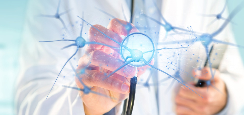
Understanding the nervous system allows you to better understand how the body works, how muscles work, how to recover, etc. . . and thus contribute to the well-being and health of your body.
Neurons are the cells that constitute the basic functioning of the nervous system. They allow the transmission of influxes, also known as nerve messages.
Its physiology is composed of a cell body that contains the cytoplasm, a nucleus and two extensions: the axon and the dendrites. The different types of nerve cells are divided according to their cytoplasmic extensions.
The dendrites are the shorter and more branched extensions; their role is to ensure the reception of nerve messages. And the axon, the second extension is unique and very long, its length can exceed one meter.
This atonic extension of the neuron is characterised by a terminal arborisation with numerous branches covered with synaptic buttons. It is through these boutons that the neurons transmit nerve messages to another neuron or to a muscle.
By releasing chemical transmitters, the receptor will be able to react to the input. The axons are also covered by a myelin sheath that allows them to be isolated and a Shawn sheath.
The fact that axons are covered by the myelin sheath ensures a faster transmission of nerve messages to the receptors.
NERVOUS SYSTEM ROLE
Humans are equipped with several billion neurons. Each one has an important role in the functioning of the nervous system. Therefore, neurons can be classified according to their function.
A distinction is made between:
- sensory neurons: this is a category of neurons which transport impulses from receptors to the central nervous system.
- efferent neurons: these are the cells that are responsible for transmitting information from the central nervous system to the receptors: muscles or glands. As soon as the message is received, the latter can respond.
- Association neurons are located in the nervous system. They complete the network of neurons, as they are responsible for the communication between sensory and motor neurons.
- Nerves: are the group of axons wrapped in connective tissue.
- Ganglia: are the collection of neuron cells that lie outside the central nervous system.
The basis of the relationship between nerves and muscles
Everything that is related to command and information emanates from the brain and spinal cord. After receiving messages, the muscles manifest themselves by performing actions as a response.
In order to provide each piece of information, myelin ensures that ion exchange is limited at the level of the myelinated fibres. These actions are manifested in the nodes of Ranvier with rapid action towards the next: this is saltatory conduction. The thicker the myelin sheath, the faster the ion exchange from the nodes of Ranvier to another.
The presence of the synapse at this level of transmission will allow messages to be delivered to the receptors. The space between the nerve tissue will be filled by this region of interaction.
The flow of impulses is provided by the chemical neurotransmitter molecule acetylcholine. The generation and propagation of a potential action on the surface of the muscle fibre allows the release of calcium from the reticulum.
The latter will travel to the interior of the muscle cell to receive the information. The command made by the neuron in the spinal cord will thus be properly transmitted to the skeletal muscles.
Motor neurons are responsible for controlling muscle movements. They function like any other cell with a nucleus and extensions, but it is because of their structure that they can perform their transmission role. They are composed of:
- a cell body containing the nucleus and made by dendrites. They are located in the spinal cord, in the grey matter.
- an axon that extends to the skeletal muscles and is covered by a myelin sheath
- terminal branches that bind to muscle fibres in the motor plate or neuromuscular synapse.
As a motor unit, all branches from the same motor neuron respond uniformly to the message sent by that same motor neuron. This means that a motor unit is independent and can perform an action in response to stimulation.
The looped network in neurons
The network of neurons is simple due to a non-looped circuit. However, the arrangement of the nerve fibres in a given circuit can result in different types of response. This is the case in a looped neural network.
A looped circuit is the result of a synapse between a collateral branch of the second neuron with an association neuron which in turn synapses with the first neuron in the chain.
Responses thus depend on the arrangement of nerve fibres in the neuron networks. In a looped network, there can be a transformation from a simple device to a complex combination that can generate different kinds of behaviour.
This means that the messages received will generate a response according to another logic, the result of an interconnection between neural chains with association neurons.
The capacity of a muscle to respond to neural information
To understand the capacity of a muscle, we must analyse its performance in relation to the work required. This allows us to know the response of a muscle at a given moment in time, according to the command given.
The question that arises is: how can we determine the number of additional motor units that need to be activated to lift a 200 kg load rather than a 100 kg load?
To get the answer to this question, we need to analyse the activity of the neuromuscular spindles. This is equivalent to seeing the contraction of the structures attached to the muscle envelope when the voluntary muscle contracts.
There is a difference between intrafusal muscle fibres and other muscle fibres. Intrafusal muscle fibres do not contain myofilaments, but numerous nuclei which are at the centre of the primary or annulo-spiral endings.
When stretched, the fibres deform into an anisometric and isotonic contraction that maintains a low and consistent tension at the cells.
The important thing to remember is that these fibres perform the same movements as the others: contraction and stretching, but with a constant and low tension, nerve endings are not very stimulated.
In the case where there is a greater resistance to the contraction which becomes more and more isometric, the tension as well as the fusorial fibres stretching will increase and transmit numerous information to the central nervous system.
In response to these different impulses, the latter will increase the muscular tension until an adequate force is reached to allow the effort or restrict the contraction. If the resistance is too great, the contraction can cause injury.
Passive stretching of the fusorial fibres is also possible through a reflex contraction called the myotatic reflex. If the muscles are overstretched, the proprioceptive receptors in the tendons will respond by inhibiting the contraction. The myotatic reflex plays a protective role here and can be considered a postural reflex.
The response to nerve information is especially dependent on the fusorial fibres and proprioceptive receptors in the tendons. These two elements ensure an isometric contraction that guarantees the tonicity of the muscles and the control of the posture.
Structures involved during reflexes
The appropriate response to information from the central nervous system is the reflex. In general, stimuli cause reflex actions in the internal mechanisms of the body. In the example of a change in body temperature, the body will act accordingly.
This means that the information goes up to the nervous system and forces the hypothalamic thermal regulation centre to set up a mechanism that will control the temperature until it reaches normal. There are many reflex reactions to a stimulus around the human body.
The sensation of burning will cause an immediate withdrawal of the hand from the source of the fire.
Apart from these innate reflexes which are triggered by the presence of a stimulus, there are also acquired reflexes which require learning. Walking, driving or working on a computer keyboard, for example, are learned reflexes, because they have to be learned little by little in order to be mastered.
Thanks to learning, some movements no longer require conscious intervention. Just start and the rhythm continues, such as putting a spoon in the mouth to eat. For 2 year olds, performing this action requires an enormous effort and greater concentration. Through repetition and education, certain actions are integrated into the nerve pathways and become automatic.
In order to have a reflex, acquired or innate, the succession of 4 phenomena must be respected, namely:
- reception: by the fusorial endings
- transmission: through the spinal cord and sensory neurons
- integration: through the central nervous system
- execution: by motor neurons and muscles
The reflex arc
The best known reflex arc is the patellar or patellar arc, often practiced by doctors, which involves two groups of neurons.
When the doctor sends a percussion on the patient's knee, the patellar tendon of the quadriceps femoris stretches the muscles. When this happens, the receptors specialised in the muscles, the neuromuscular spindles, send a message via the sensory neurons to the spinal cord. A synapse with motor neurons occurs there to ensure the continuity of the information.
At this point, the spinal cord responds and sends a response to the quadriceps which causes the contraction: the leg lifts. Both sensory and motor neurons are involved in this type of reflex, but there is only one synapse that occurs, it is a monosynaptic reflex.
As the patellar reflex is part of the reflex pathways, it also follows 4 functional phases:
- the reception provided by the fusorial endings which are sensitive to stretching
- transmission or afferent pathway by the sensory neurons which transfer the messages to the spinal cord
- the integration by the central nervous system at the synapses of the spinal cord
- the efferent pathway through the motor neurons which transmits the response to the effector organ or the muscle.
In the case of the withdrawal reflex, there are 3 groups of neurons and two synapses involved, which is why it is called a multi-synaptic reflex. When the hand feels a burning sensation, the withdrawal is instantaneous.
This action is the consequence of the entry into force of pain-sensitive receptors called nociceptors, dendrites of sensory neurons and synapses between association neurons.
First there is the reception, then the transmission of the message by the neurons which synapse with interneurons, then the integration by the spinal cord and finally the contraction of the muscles of the hand and arm caused by the motor neurons.
At the same time, there is reciprocal resistance from the neurons that block the antagonistic muscles.
There are also cases where the reflex does not require the intervention of the brain. These situations are the result of a conscious inhibition or facilitation of reflex actions.
For a baby, emptying the bladder is a reflex in which the brain is not present. When the bladder is full, it simply empties. While growing, a child learns to control the bladder. At a critical threshold, the bladder sends a signal to the nervous system and the nervous system responds to avoid the facilitation reflex.
The development of a conscious resistance to the urination reflex also provides this control.
Conscious inhibition can be learned and allows humans to think before they remove their hand when they get burned. Consciously you do not withdraw your hands if you are carrying a very hot object while someone is near you.
You hold the object until the other person is safe even though the response to the stimulus requires you to immediately withdraw your hands. Athletes often practice this conscious inhibition during training. It is difficult, but with perseverance and repetition, despite the pain, the body gets used to it.
The automatic movement
Automatic movement is the consequence of learning reflexes. A motorist will stop his car when he is at a crossroads and the traffic lights are red. It is a repeated execution of gestures in a mechanical way without the intervention of thought. The functioning of the nervous system is based on simple reaction steps.
In this example, the reception is the observation that the traffic light has turned red. This information is received by the eyes and thanks to the sensory fibres, it is sent to the central nervous system to obtain a response.
It is during integration that the central nervous system analyses, classifies and interprets the information. At the end of this phase, the appropriate motor neurons are excited and the response is sent to the muscles involved.
The input received will be interpreted by the removal of the foot from the accelerator and a pressure on the brake pedal: the effectuation.
As mentioned above, all the responses that require the intervention of the brain are based on 4 phases: reception, transmission, integration and execution. The information is transmitted to the receptors in a sequential manner: first the information travels to interneurons in the CNS and then the response is transmitted by motor neurons.
To allow the transmission of information and reflexes, the nervous system contains millions of sequences of neuronal elements: this is the nervous circuit. This circuit is ensured by the synapse: the contact of a neuron, thanks to the axon, with dendrites or a cell body of another neuron.
The synaptic cleft conditions the contacts between neurons. In this connection, it is essential to note the difference between pre-synaptic and post-synaptic neurons. Pre-synaptic neurons are those with an axon ending at a specific synapse and post-synaptic neurons have a dense membrane anchoring the receptors to which neurotransmitters bind.
The control of voluntary movement and posture
Muscle actions are the consequences of electrical stimulation at the cortical surface. The neurons that control movement in the face, hand and torso are different. In the torso, the neurons of the primary motor area are involved, whereas in the face, the neurons of the base area are involved. With more precise movements of one or more muscles, the cortical surface area for stimulation is larger.
The neurons involved in the face and hand are much more numerous in order to provide more precise information, which is not the case in the torso.
In the programming, planning, control and execution of a gesture or voluntary movement, the involvement of motor areas adjacent to the primary area is necessary.
In the depth of the cerebral hemispheres, the basal ganglia are also important elements to consider when it comes to voluntary movement.
Responsible for precise and controlled movements is the neurotransmitter dopamine, which plays an essential role in motor function. Its absence in the case of a kinked nucleus is marked by jerky and hesitant movements.
The pyramidal and extrapyramidal pathways that transfer the response from the medulla to the motor neurons are also very important.
They ensure precise and skillful movements (pyramidal bundles) and various body postures (extrapyramidal bundles).


