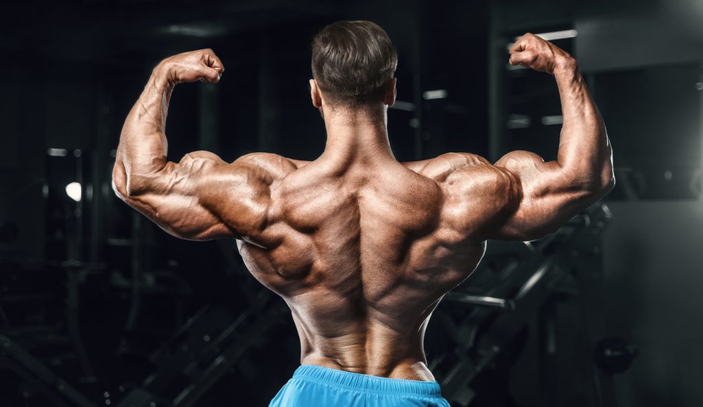
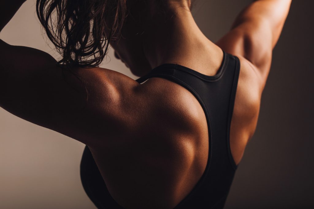
Anatomy of The Back
Many muscular groups make up the back. Among the fundamentals are the large dorsal muscles, the trapezius muscles and the lumbar muscles. Each differs primarily in shape. For example, the large dorsal muscles are intended to be larger. The trapezius muscles are located at the top of the back. They are made up of 3 fascicles, namely:
- the upper fascicle,
- the middle fascicle,
- the lower bundle
Opposite the trapezius muscles, i. e. in the lower back, are the lumbar muscles.
The lumbar spine
Since it covers the entire back, the lumbar spine is the largest muscle in the human body.
From an aesthetic point of view, the lats is an important muscle. Indeed, it is to it that the back owes its V-shaped appearance. The strength of the Latissimus dorsi also helps to maintain a good posture. This muscle connects the trunk to the forearm at the same height as the pectoralis major. It also connects the upper part of the torso to the spine and the lower part to the pelvis via a large, flat mass of tendon called the lumbosacral fascia.
Another point to remember is that the latissimus dorsi is a chest straightening muscle. It raises the shoulders and pulls them back.
The Latissimus dorsi ends at the kidneys in a fibrous blade. As an accessory inspiratory muscle, it allows the arms to be positioned upwards. This attribution of the Latissimus dorsi is simply due to the fact that it is located on the inner side of the last four ribs. In addition, there are the lower digits of the oblique abdominal muscles.
You will also notice it during certain movements: when you lower your shoulders and bring them back, this muscle brings the shoulder blades together, not to mention the extension of the dorsolumbar spine.
The Latissimus dorsi muscle is also known to lower the arm. Indeed, when you raise your arm, without this muscle, it will automatically fall back down. When you lower your arm, the Latissimus dorsi muscle can bring it back, promoting an extension movement.
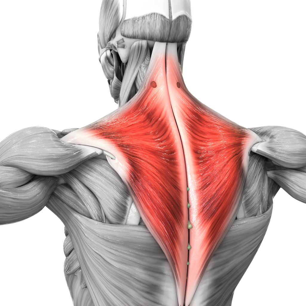
The trapezius muscle
It is on the upper part of the back that the trapezius muscle is located. More precisely, it extends from the back of the neck, at the 12th dorsal to the shoulder blade. Thanks to the trapezius muscle, the latter can rise, fall, move closer to the spine or even tilt.
The large muscle
The Larger Round muscle is inserted at three points:
- on the inner edge of the scapula,
- at the top, outside,
- at the level of the humerus
Together, the great circle and the humerus allow some movement of the back.
The rhomboid
The rhomboid begins at the medial edge of the scapula and extends upward medially to the spinous processes of the vertebrae. It inserts onto the vertebrae from the 7th cervical to the 4th dorsal.
The rhomboid attaches the upper angle of the scapula to the ribs. A lack of tone in this muscle leads to scapular detachment. On the other hand, the lateral lowering of the arm is the result of a simultaneous action of the great circle and the rhomboid.
In case the Larger Round contracts in isolation, it will rotate the scapula, moving the lower angle of the latter away from the spine.
Little round and infraspinatus
These muscles originate on the posterior part of the scapula, close to the greater tuberosity, and end very high on the humerus. They have a small part in the lowering of the arm.
On the other hand, they are necessary to perform certain movements, such as external rotation of the arm and shoulder articulation. In these movements, the lesser arm and the infraspinatus are assisted by the long portion of the triceps. The latter resists downward dislocation of the shoulder and stretches to elicit the action of the greater dorsal and greater round muscles.
Lumbar region
The lumbar vertebrae are located on the lower part of the back. They are attached to the hip. They are monoarticular muscles which play two motor roles.
On the one hand, the lumbar muscles are responsible for hip flexion. They also promote mobility of the lumbar spine. The latter has articulatory properties. On the other hand, they act as extensor and flexor muscles of the spine. The lumbar muscles are used when you lower yourself forwards, backwards or sideways.
Sometimes these muscles are also involved in rotation movements.
The spinal extensors
They consist of three columns of muscles which run parallel to the spine. These are the protruding muscles on each side of the spine.
The three vertical columns of spinal extensors attach:
- the first one at the top of the hip, on the posterior part of the sacrum;
- the second along the last lumbar vertebrae;
- the last one, the most external, is implanted on the ribs.
The middle column, formed by the long dorsal, is implanted on the dorsal and cervical vertebrae and on the skull. The innermost spine is attached to the spinous processes of the first thoracic vertebrae and to the skull. It is made up of the semispinatus and the spinatus.
When the two sides tighten, the spinal extensors arch the spine and head. But when the extensors tighten individually on the left or right, the spine bends to the same side.
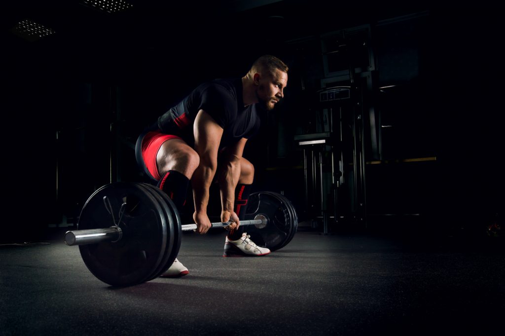
Some exercises targeting the back muscles
- The deadlift
- Successful deadlift
- Exercises to strengthen your back
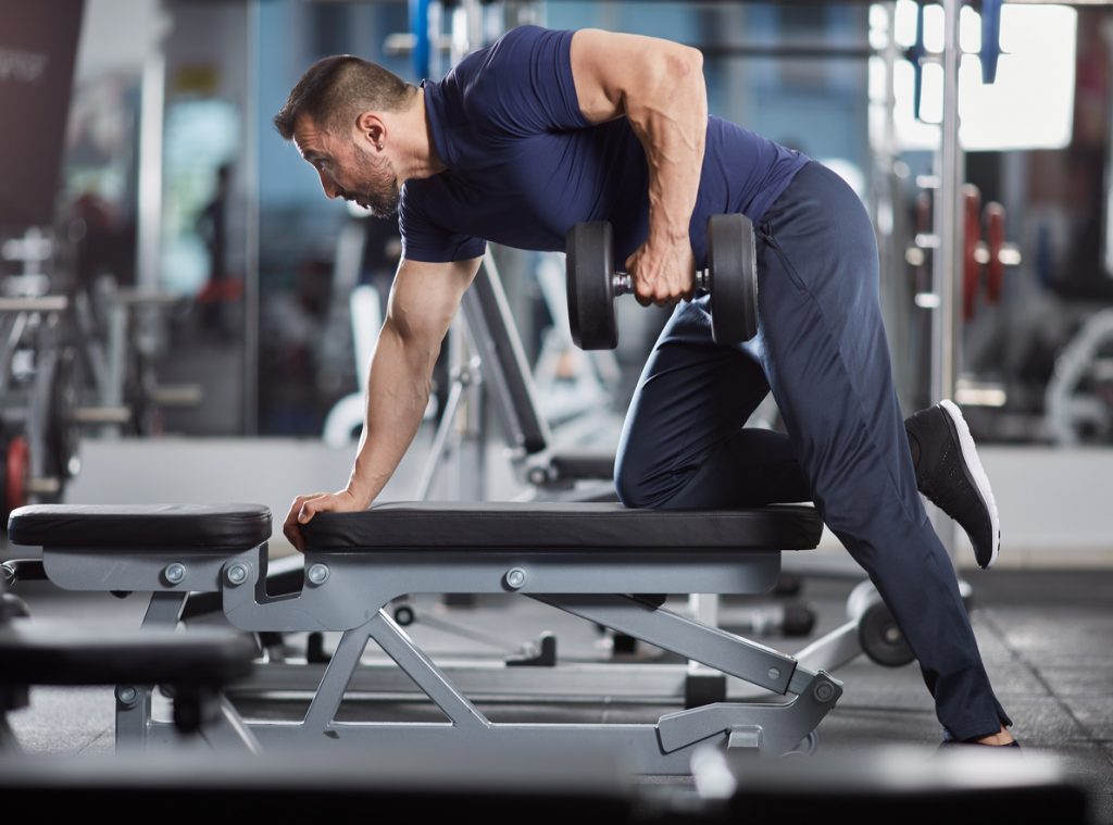
- The rowing bar
- The barbell rowing
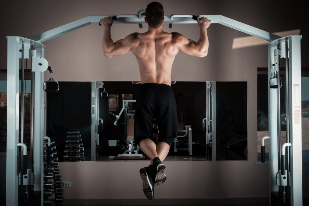
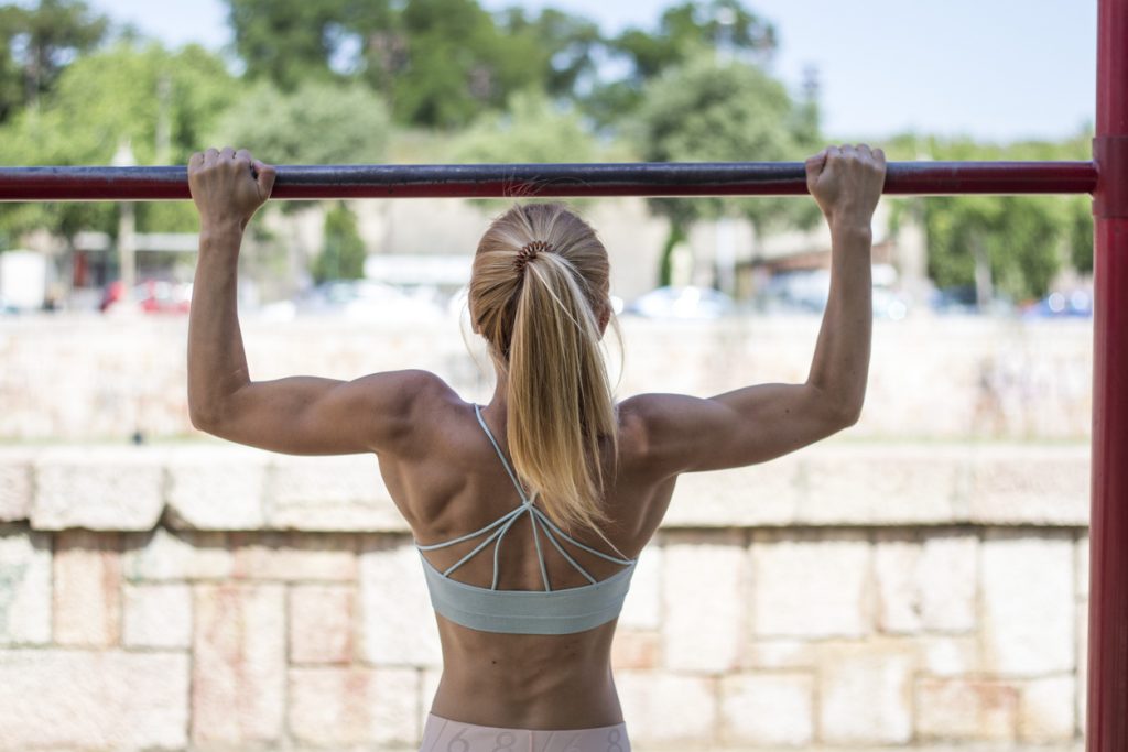
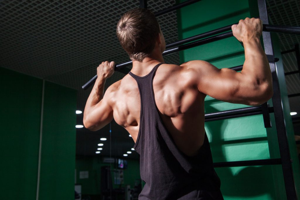
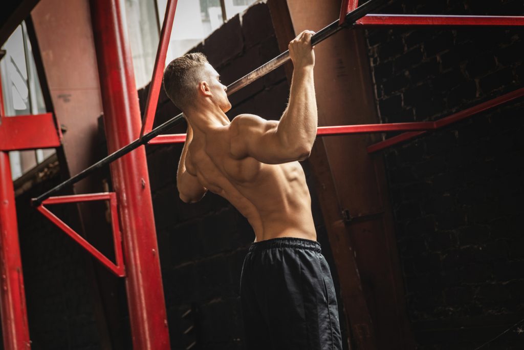
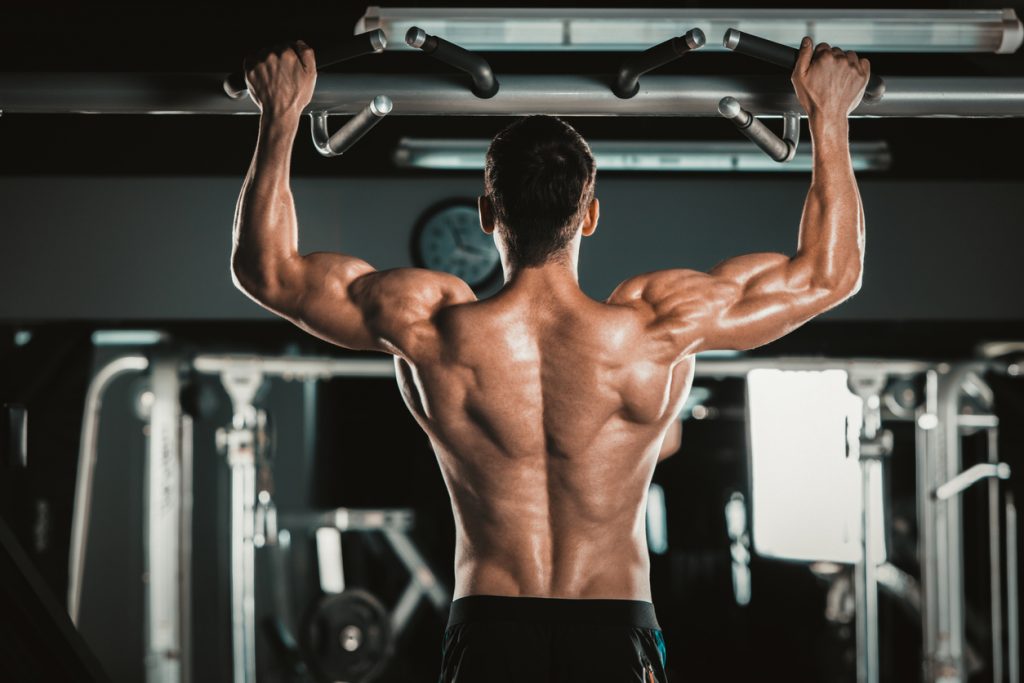
- Pull-ups
- Scapular pull-ups
- High bar pull-ups for a V-shaped back
- Exercises to increase back width
- The best exercises for the back
- Developing your back muscles