Anatomy The Buttocks
The gluteal muscles (attached to the leg) are the following:
- the gluteus maximus,
- the gluteus medius,
- the small buttocks,
- the piriformis muscle,
- obturator internus muscle,
- upper and lower twins,
- femoral square,
- iliopsoas muscle,
The gluteus maximus
Of all the gluteal muscles, the gluteus maximus remains by far the most superficial and the most voluminous. It is easily recognised by its thickness and its quadrilateral shape.
The gluteus maximus overlaps all other gluteal muscles. This explains why bodybuilders are so keen to emphasise it on the competition stage. In fact, it is the muscle fibre bundles of the gluteus maximus that are visible as striations on the buttocks of these still dry bodybuilders.
The gluteus maximus is attached to the posterior fifth of the iliac crest, the posterior part of the iliac fossa, the sacrum and the coccyx.
It is attached:
- on one side by deep fibres on a tendon;
- on another side by superficial fibres located behind the fascia lata fascia.
When you look at a mirror, you will notice that the lower edge of this muscle (the gluteus maximus) takes the form of a visible fold between the buttocks and the back of the thigh.
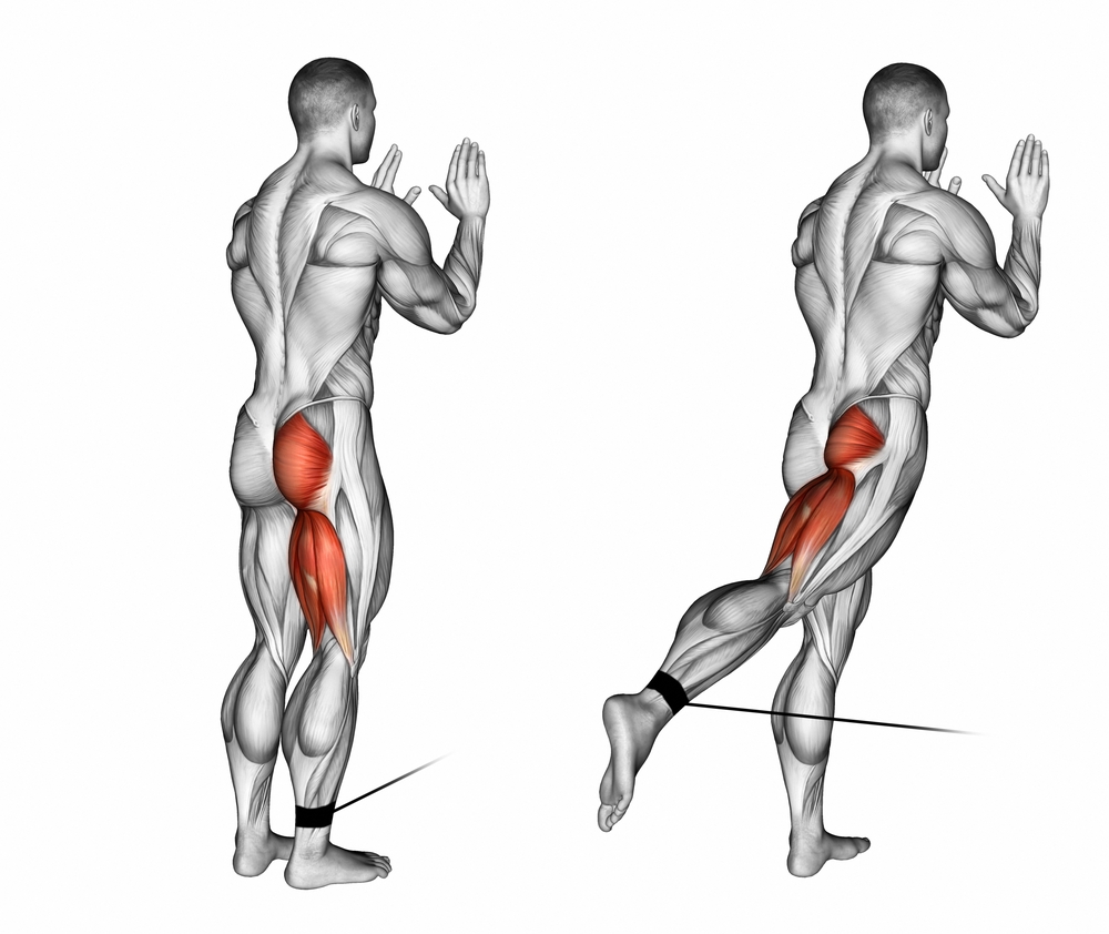
Overall, the gluteus maximus is responsible for the extension of the thigh as well as the external rotation of the thigh. In concrete terms, here are 3 fundamental roles of this muscle:
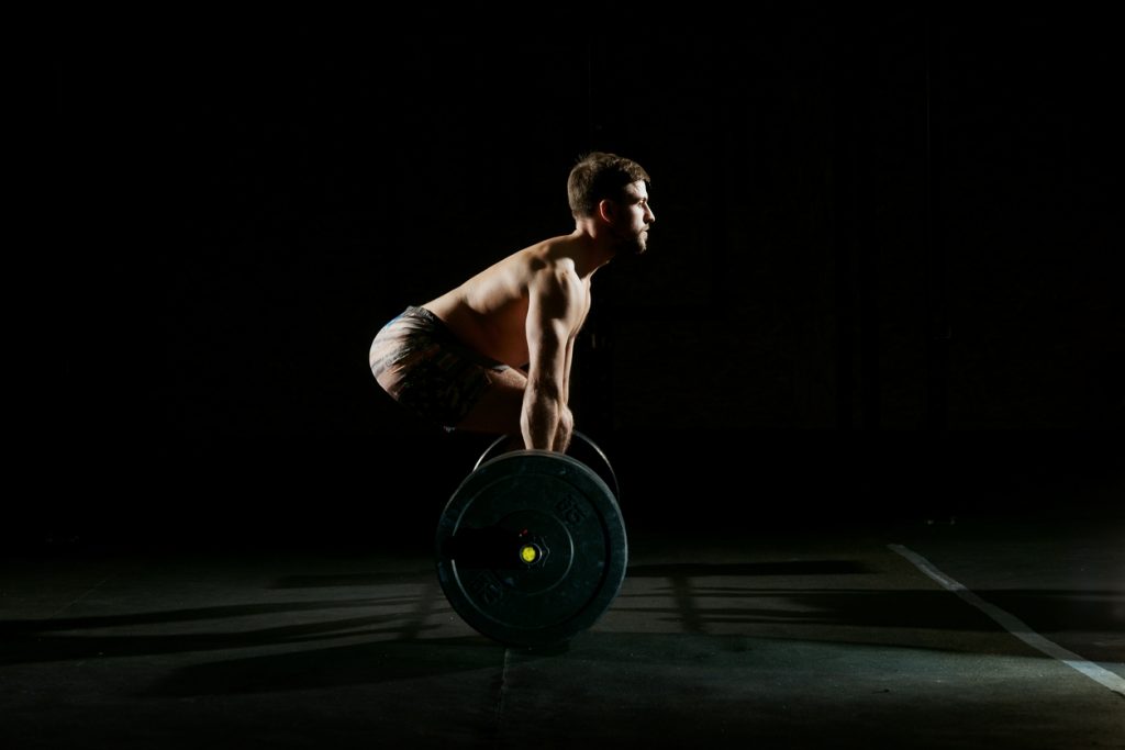
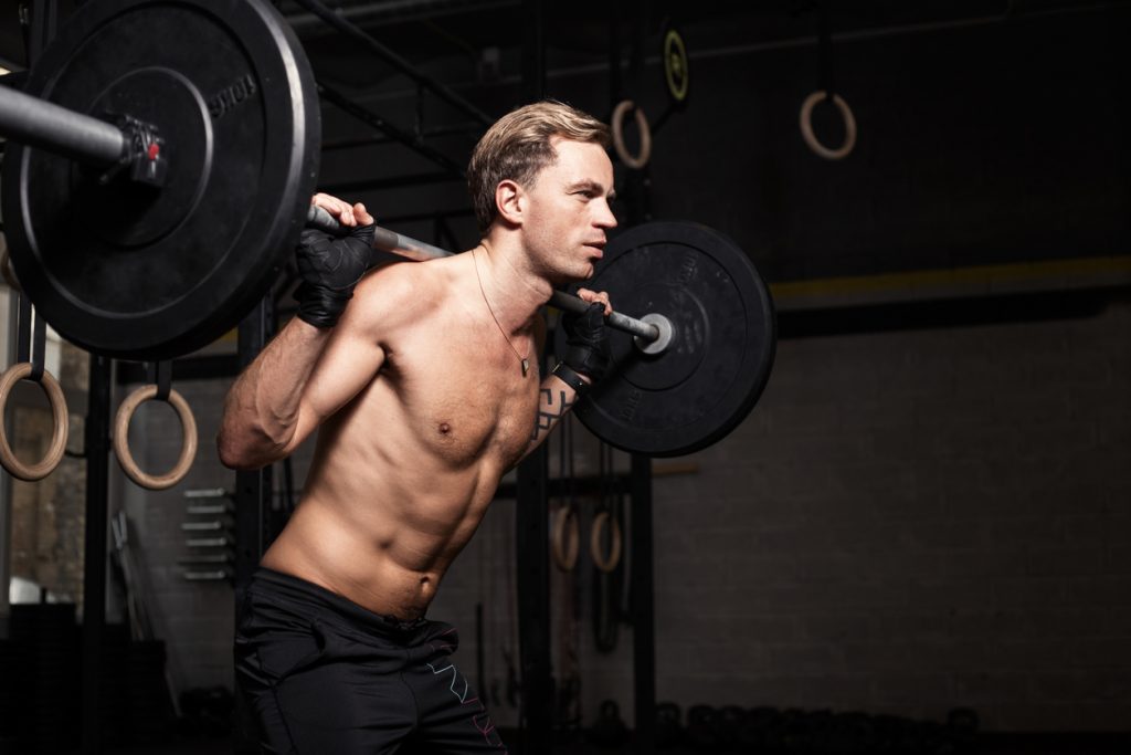
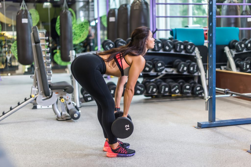
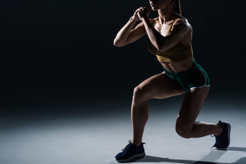
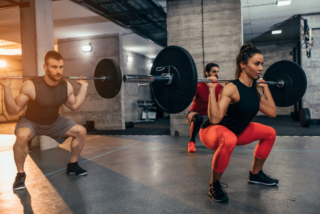
- during squat and hack squat sessions or when you perform lunges with bars or dumbbells, this muscle allows the extension of the thighs at the hip joint. It also performs a slight external rotation at the thigh.
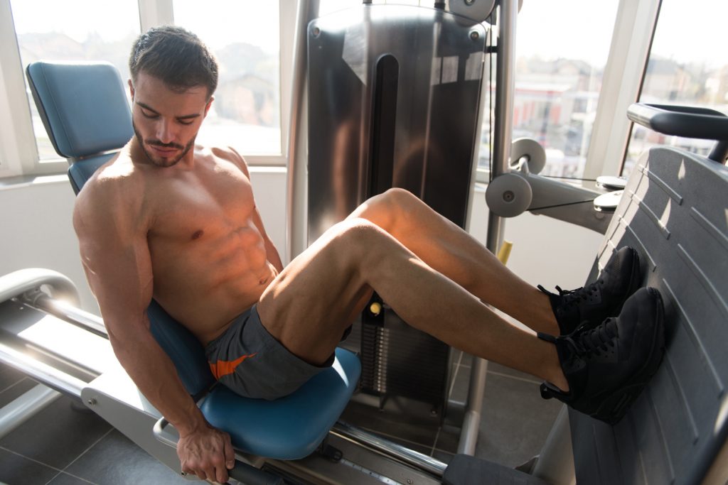
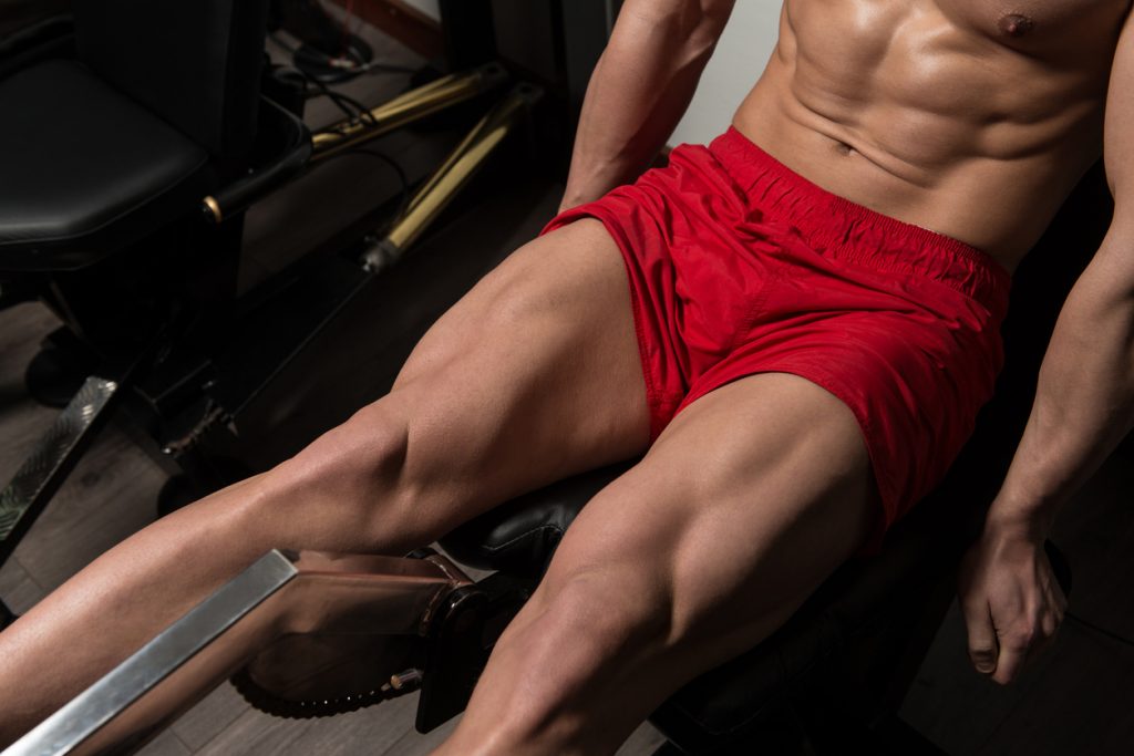
- when you do exercises with a thigh press and these require strength, this muscle contracts strongly;
It should be noted that through these exercises, which are basic movements, other muscles of the lower limb interact with the work of the gluteus maximus. However, this muscle is not used much during the walking sessions. It is even relaxed when you are standing still.
The gluteus medius
The gluteus medius is triangular in shape and is essentially covered by the gluteus maximus. You will find it at the inner three quarters of the iliac crest and the anterior and superior iliac spine. It ends in a tendon that is in turn attached to the outer surface of the femur.
The upper part of the gluteus medius muscle is not hidden in any way under the gluteus maximus. In any case, this muscle remains invisible in bodybuilders.
The gluteus medius muscle has two basic roles:
- adductor,
- motor.
The gluteus medius muscle as an adductor
To gain passage in leg thrusts, especially in the lateral position, you will need the gluteus medius muscle. The same is true when you exercise on machines to work your hip and thighs or when you perform abduction movements on a low pulley.
The motor role of the gluteus medius muscle
When you take a step forward, the left gluteus medius pulls the left side of your pelvis down, allowing you to perform all walking movements with ease.
That's not all! This muscle also allows your right foot to leave the ground when you walk, without collapsing.
The gluteus maximus
The gluteus minimus muscle is located just below the gluteus medius, above and medial to the external iliac fossa. Its triangular shape makes it easily recognisable, especially as it ends in a tendon attached to the anterior aspect of the femur.
The gluteus minimus has more or less the same insertions as the gluteus medius. This is the reason why these two muscles perform the same functions (rotation and walking).
More precisely, the gluteus minimus muscle contains internal and posterior fascicles which perform the roles of internal and external rotation respectively.
The motor role is derived from the fact that the gluteus maximus provides pelvic tilt. This allows the opposite foot to leave the ground and rise when you walk.
The piriformis muscle
Formerly known as the pyramidal muscle of the pelvis, the piriformis muscle also represents a triangle. It has several specific characteristics. You can recognise it quickly and easily. Here are some of them:
- the piriformis muscle is located deep in the body;
- it is a narrow muscle that extends from the sacrum to the greater trochanter;
- on the mirror, its upper edge represents the line between the two dimples;
The piriformis muscle acts as:
- external rotator when the thigh is tense.
- Abductor when the thigh is flexed.
Here is a concrete example: the piriformis muscle is used when you are sitting and doing exercises on an abductor machine.
The obturator internus muscle
The obturator internus muscle is easily recognisable because it is located under the piriformis muscle. It owes its name to its location, or more precisely to its insertion. It is located near the hole in the hip bone. The latter is also known as the obturator foramen.
Ending on a tendon located in the digital fossa, the obturator internus muscle performs the same functions as the piriformis muscle. In other words, it acts as both an external rotator and an abductor.
The upper and lower twins
The upper and lower twins are located under the piriformis muscle and are distinguished by their tapered shape. They enclose the obturator internus muscle and the muscle fibres in the surrounding areas.
The upper gastrocnemius is grafted onto the lateral aspect of the sciatic spine. The inferior gastrocnemius is located on the lower edge of the sciatic notch, near the ischial tuberosity. Both have the same termination, i. e. at the level of the digital fossa on the medial side of the femur.
Together, the upper and lower twins hold the head of the femur in the acetabulum of the hip bone. This is how the body can ensure that the hip is stable when you perform a certain movement. It can even be said that these two muscles play the same role as the piriformis and obturator internus muscles.
The femoral square
Have you touched a flat, short, rectangular muscle? It must be the quadratus femoris muscle, formerly called quadratus femoris. Located under the obturator internus and the twins, it is inserted at the level of the external edge of the ischial tuberosity. Its muscle fibres terminate on the lower part of the posterior intertrochanteric line of the femur.
The quadratus femoris provides external rotation of the thigh. It also holds the femoral head in the hip joint.
The iliopsoas muscle
Senior scientists know this muscle mainly as the psoas iliacus muscle. It is found on the upper part of the inner side of the femur.
As the name suggests, this muscle is made up of two entities: the iliacus muscle, which fills the entire iliac fossa, and the psoas. The latter extends from the 12th dorsal vertebra to the 5th lumbar vertebra.
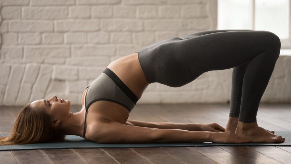
The particularity of the iliopsoas muscle lies in its strength. Indeed, the ilio-psoas muscle is at the origin of hip flexion. It also tilts the pelvis into anteversion, thus deepening the lower back. This muscle is used twice when you do abdominal exercises.
To give you an idea of its importance, here are some points to remember:
- when the ilio-psoas muscle contracts on one side only, a hollow appears on the lower back and the spine in a scoliotic attitude;
- when the iliopsoas muscle contracts on both sides, the thighs bend over the trunk (or vice versa) at a fixed point.
A little clarification: when you bend your legs during your abdominal sessions, you encourage the shortening of the iliopsoas muscle. Generally, this is what causes pinching and shearing of the lumbar discs and cramps when standing.
In order for the iliopsoas muscle to perform its functions fully, it must meet certain criteria. For example, a long muscle is essential for a good night's sleep and for lengthening the legs. If you are a keen walker, your iliopsoas muscle must be sufficiently stretchy to allow you to take long steps.
If you want to stretch your iliopsoas muscle, lunges are your best bet. Indeed, the exercise to do is simple: you extend your back legs, then you tighten your buttocks and your abdominals so as to create a hollow on the lower back.