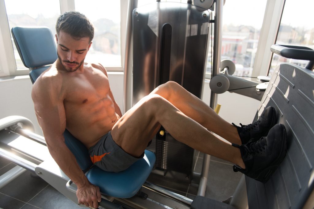
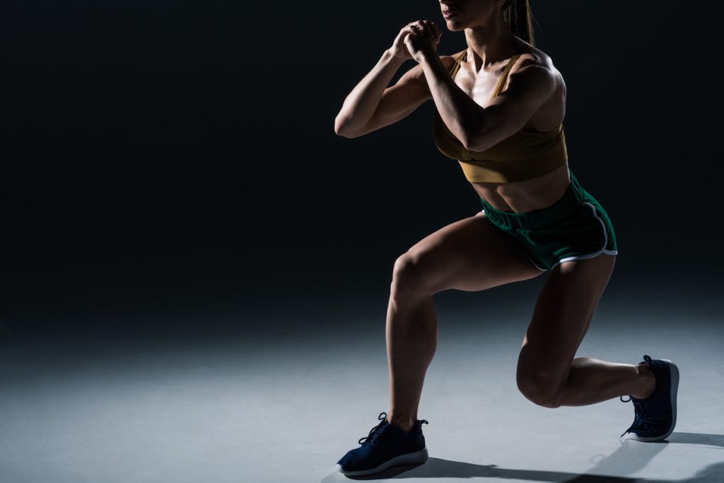
During strength training, this part of the body is often neglected because it is less visible than the upper part. And we think that this practice on the Ischios is optional. However, in addition to the enormous impact on the general aesthetics of your form, not having the leg muscles well developed can promote major drawbacks on your health.
We offer you all the keys to building up your hamstrings in this feature. Starting by giving anatomical explanations to better understand how these muscles work, then comes the study of the best strength training exercises (with execution tips, as most exercises for the hamstrings involve the pelvis and lumbar, so they are not to be taken lightly), but also several training programs, to include in your thigh training, to effectively muscle this muscle group.
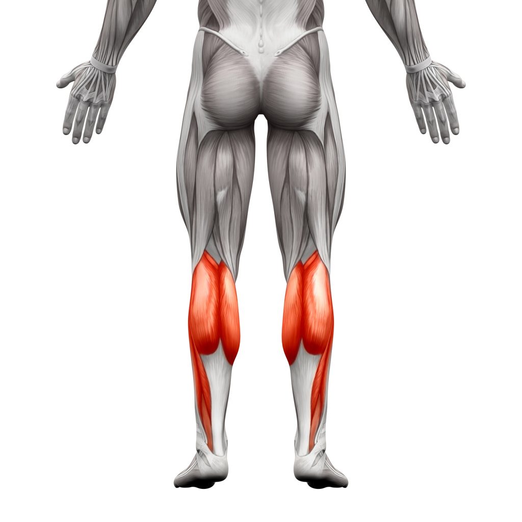
Anatomy of the leg muscles
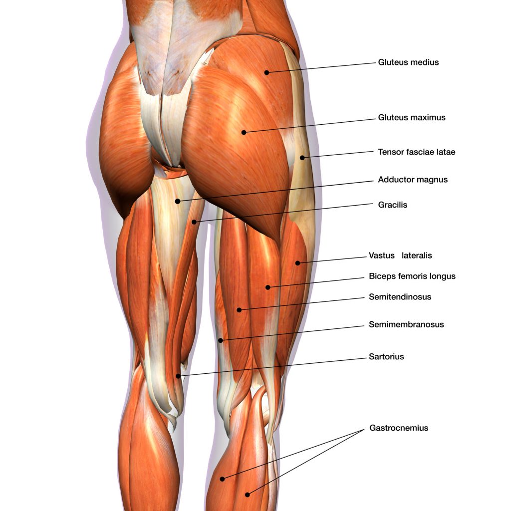
Consisting of the biceps femoris, semitendinosus and semimembranosus, the muscles work together at the hip and knee. The hamstrings are involved in the movement of the knee and are involved in the action of walking and standing.
-
The Biceps femoris
This is the external muscle of the posterior thigh. It has two heads: the first is bi-articular and is the long portion, the second is mono-articular and is the short portion. These portions are located respectively at the level of the coxal bone at the fibula (first) and the femur at the fibula (second). As far as insertion is concerned, the long portion has a common part with the ½ membrane and ½ tendon, the lower part of the ischial tuberosity, behind the adductors.
The short portion is inserted at the level of the lower ½ of the outer lip of the linea alba, below the gluteus maximus. The Biceps femoris is characterised from the Oblique downwards. Indeed, the long portion passes medial and posterior to the short portion. This muscle is a hip extensor, it also locks the knee in extension. In other words, this part of the leg allows the knee to flex and the tibia to rotate externally on the femur. You should probably know that the sciatic nerve is located between the two heads. There is also a common fibular nerve that follows the terminal tendon to the head of the fibula.
-
The semi-membranous
This is the second component of the Ischio leg muscles, it is composed of articular Bi, located in the posterior compartment of the thigh internally, between the coxal bone (ischium) and the tibia. The muscle has a common part with the biceps and ½ tendon, the lower part of the ischial tuberosity, behind the adductors. This membrane runs vertically down the medial and posterior part of the thigh and ends in 3 tendons:
- Direct: at the posterior aspect of the upper end of the tibia
- Reflex: below the medial tibial plateau
- Recurrent: fabella, this is the oblique popliteal ligament
Concerning its innervation, the semimembranosus is innervated by a branch from the sciatic nerve. This bundle is common to the ½ membrane and the inferior head of the adductor magnus. This part of the leg is also a hip extensor, with the knee locked in extension. It allows for knee flexion and rotation of the tibia on the femur. The reflected tendon slides under the tibial collateral ligament.
-
The semitendinosus or crow's feet muscles
This is a set of 3 bi-articular muscles of the thigh: the gracilis, the Sartorius and the semitendinosus. Anatomically, these 3 muscles are stretched from the hip bone to the tibia and have a different insertion, a different innervation but a common insertion on the medial side of the tibia: the crest of the crow's feet.
The Sartorius is situated on the superficial plane of the anterior compartment. The Gracilis is located in the medial compartment. Finally, the semitendinosus is located in the posterolateral compartment.
The Gracilis inserts at the level of the medial 1/3 of the inferior border of the ischio-pubic ramus; the Sartorius is integrated on the lateral face of the anterosuperior iliac spine in front of the tensor fascia lata.
The semitendinosus is devoted to the inferior part of the ischial tuberosity, behind the adductors, in common with the biceps femoris and the semimembranosus.
Regarding the course, the Gracilis is oblique downwards and outwards, and descends medially and posteriorly from the medial condyle towards the crow's feet. The Sartorius runs up and down, out and in, front and back, in an S-shape.
Finally, the semitendinosus runs vertically down the posterior side of the semimembranosus and passes behind the medial condyle of the femur. The tendon on the upper 1/3 of the medial aspect of the tibia is called the crest of the crow's feet.
These 3 sets have a common action, which is the internal rotation of the thigh, the bending of the leg on the thigh. There is even an active medial knee joint ligament, which helps prevent medial sprains due to the medial collateral ligament they support.
But the 3 components also have various functions, such as flexing the hip and knee, rotating the tibia on the femur, controlling stability for the Sartorius. It is the muscle of the cross-legged position.
As for the Gracilis, it is responsible for flexion of the leg on the thigh, internal rotation of the knee and active ligament of the knee, and it is an adductor of the thigh on the pelvis. The semitendinosus are extensors of the thigh/pelvis and flexors of the leg/thigh, but also medial rotators of the leg.
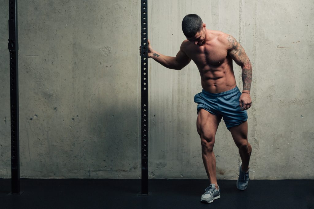
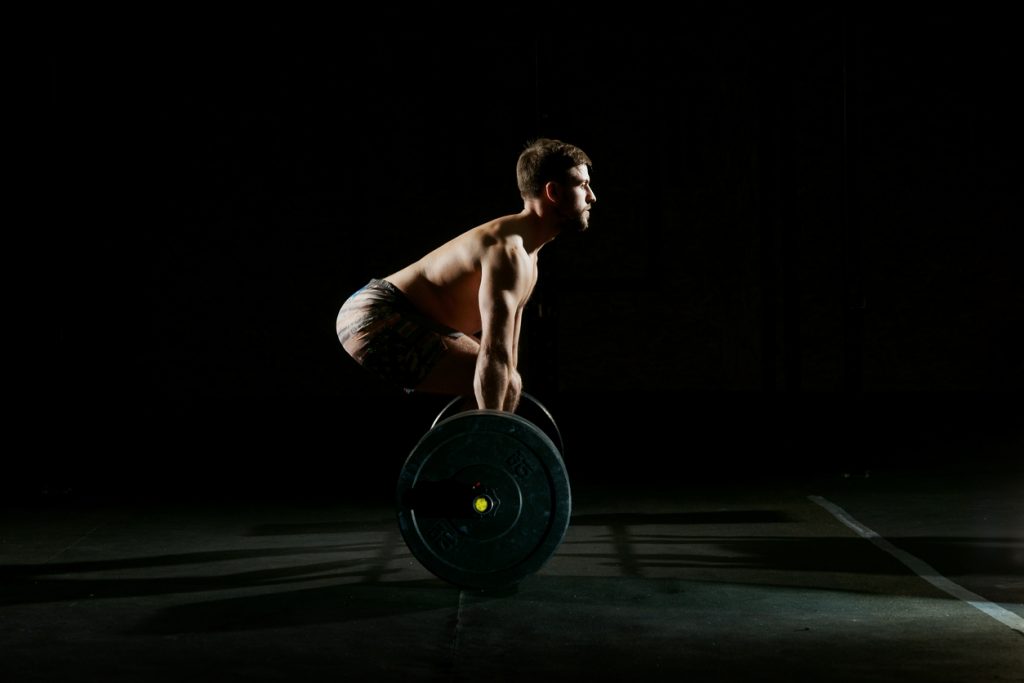
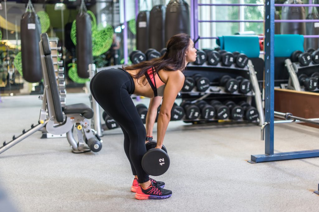
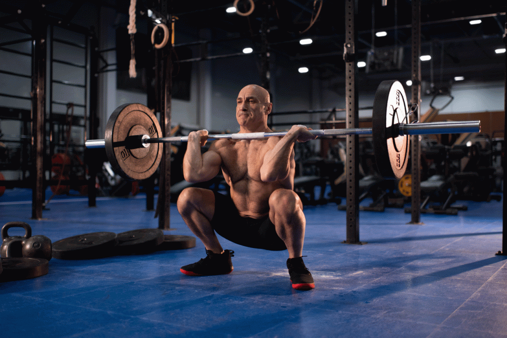
Leg exercises: The Good morning
The "good morning" exercise uses the same muscles as the deadlift, but is more dangerous due to the position of the bar on the trapezius muscles.
Execution :
Stand with your feet firmly on the floor, one shoulder width apart. The bar is placed on the trapezius muscles.
Bend the chest forward until it is horizontal, keeping the back straight.
Stay sheathed and then return to the starting position keeping the legs straight or slightly bent.